
preclinical imaging
About Us
The Laboratory of Imaging for small animals of IBSBC -CNR is part of Multi-Modal Molecular Imaging Italian (MMMI) node of the European Research Infrastructure EuroBioImaging (EuBI).
It is located inside Nuclear Medicine Department of San Raffaele Research Institute upon a formal agreement. The services provided are possible thanks to the collaboration also with the Department of Health Sciences and TECNOMED Foundation of the University Milano – Bicocca (UNIMIB).
The research activity of the Laboratory of Imaging for small animals covers Emission Tomography based molecular Imaging, from radiopharmaceutical development to preclinical as well as clinical application. The group has expertise in radiolabelling with C-11, F-18 and Ga-68 including methods and procedures required according to EU rules. In addition, the group has developed methods and strategies for positron emitters metals isotopes binding, as Cu-64 and Zr-89. A larger number of radiopharmaceuticals has been already validated and applied at preclinical and clinical levels for the study of brain neurotransmission systems (serotonin, dopamine, benzodiazepine receptors, opiate receptors), amyloid plaques, activated microglia cells and for the in vivo imaging of biochemical pathways involved in cancer (choline kinase activity, cell proliferation, regional tissue hypoxia, glucose metabolism). The main fields of application are CNS diseases, inflammation and cancer and most of research activities are carried out in close collaboration between preclinical and clinical researchers.
Applications
Cells

Cells
We can apply screening protocols of radiolabeled compounds in cells for applications as imaging probe or therapeutic agent. Molecules of different sources as chemicals, peptide or nanoparticles/liposomes can be tested.
Animal

Animal
We have experience in preparing and monitoring of different rodent models of disease (onco, neuro, cardio, inflammation) for in vivo imaging applications (CT/PET/SPECT/OI) and ex vivo distribution and dosimetry. We are equipped for animals housing in longitudinal studies.
Microbiome
Microbiome
In vivo imaging could be applied to check microbiome composition in animal models under different conditions as gender, ageing or different treatments.
Clinical/PRECLINICAL

Clinical
We can set up proof of concept studies in animals to replicate clinical trials of imaging and treatment protocols: diagnosis using multi-modal imaging, therapy and follow up.
Nutrition

Nutrition
Imaging techniques can be applied to study the effect of different food regimens in animals. We can also evaluate the effect of natural extracts in cells and animal models of disease (onco, neuro, cardio, inflammation).
MOLECULES
Molecules
We can radiolabel and evaluate different types of molecules (chemicals, peptides, liposome/nanoparticles) as potential probes for PET/SPECT in vivo imaging. We provide also screening of molecules (or combinations) as therapy in animal and cellular models of disease.
Facility
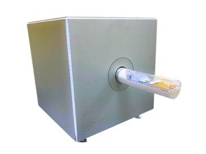
18 MeV Cyclotron (Iba)
Radiosynthesis cells and modules for radiotracers production
CT/SPECT/PET System
(Molecubes)
Cyclone Plus Storage Phosphor System (Perkin elmer)
Gamma-counter (Perkin elmer)
Ivis Lumina Serie III (Perkin elmer)
Services
The Laboratory of Imaging for small animals provides services within the European Research Infrastructure EuBI, as part of Multi-Modal Molecular Imaging Italian (MMMI) node.
Preparation and housing of different animal models of pathology in mouse and rat induced by injection of cells, toxins and chemicals. Models evaluation, validation of new radioligands and of new treatments
Labeling of established clinical-standard molecules used in nuclear medicine for the diagnosis of various diseases like cancers, cardiovascular and brain disorders (i.e. [18F]FDG, [18F]PSMA, [18F]FDOPA, [11C]methionine…). Functionalization study of different species (i.e. small molecules, peptides, proteins, nanoparticles and liposomes); introduction of PET/SPECT radioisotopes for the production of new probes for diagnosis/treatment in animals (research-standard)
Postmortem sampling of tissue and fluids deriving from animals injected with radiotracers: gamma-counter counting and radioactivity concentration quantification
Image and data analysis deriving from in vivo and ex vivo acquisitions on animals and samples: radiotracers quantification, radiotracers kinetics and different parameters evaluation. Standard analysis with commercial software or implemented analysis using customized tools
Price List
Probe design
(feasibility study)-
Cost per production* (€)
Probe labelling
(custom, small or bio-molecules)-
Cost per production* (€)
Radiosynthesis of conventional probes
-
Cost per production* (€)
PET/SPECT/CT: β-Cube, γ-Cube, X-Cube
(Molecubes, Gen)-
Cost (€/h)
Optical Imaging: Ivis Lumina III
(Perkin Elmer)-
Cost (€/h)
Ex vivo biodistribution
-
Cost (€/h)
Ex vivo autoradiography
-
Cost (€/h)
Data analysis
-
Cost (€/h)
Additional services
-
Costs of additional services will be quantified based on technology requested